Optical coherence tomography
Medical imagingWikipedia
138
MENTIONS
43
EPISODES
36
PODCASTS
Search complete. 138 mentions across 43 episodes found for "Optical coherence tomography".
Oct 28, 2026

Diana DoHOST
And when we look at diabetic macular edema, we also see a benefit.
Diana DoHOST
And, you know, maybe Michael, tell us a little bit about what biomarkers on OCT and how we can use that in our clinical practice.
Michael JavahariGUEST
Well, we need to look.
Michael JavahariGUEST
We look hard at Z-dates.

Bryce PerryHOST
And guess what? They have found a pattern among people who later went on to receive a Parkinson's diagnosis.
Bryce PerryHOST
Ready? The reveal is that an OCT eye scan can show Parkinson's-related retinal changes years before the disease is clinically diagnosed.
Bryce PerryHOST
Who knew? In the study, the differences were found in scans taken an average of about seven years before people presented clinically with Parkinson's.
Bryce PerryHOST
Seven years.
Bryce PerryHOST
You know, Parkinson's may have been leaving a clue at the back of the eye while everybody was still waiting for the hand, the foot, or the walking patterns to make the official announcement.
Bryce PerryHOST
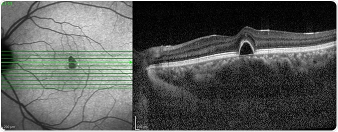
The scan is called Optical Coherence Tomography or OCT.
Bryce PerryHOST
And it uses light to create an incredibly detailed cross-section of the retina.
Bryce PerryHOST
And researchers found thinning in particular retinal air layers.

Vít PohankaCORRESPONDENT
Some of the microscopes also have built-in technology for detailed imaging of the eye's internal structures.
speaker_9SOUNDBITE_SPEAKER
The high-end technology that we're having here, so these are like the best microscopes, they have already included intraoperative OCT also, which we don't use here, but they still include it in the machine, so with the screen, with the 3D screen and the microscopes, like really high-end technology, it's like the best of the best.
Vít PohankaCORRESPONDENT
OCT, or optical coherence tomography, provides detailed images of structures inside the eye.
Vít PohankaCORRESPONDENT
Although that particular feature isn't being used here, modern microscopes offer other advantages, especially when surgeons need to work with extraordinary precision.
Vít PohankaCORRESPONDENT
Better lighting and magnification help them distinguish the delicate tissues they are operating on.

Meenal AgarwalGUEST
you know, just like we're advancing in brain health, we're starting to advance in eye health.
Meenal AgarwalGUEST
And so there are scans, one in particular called the Optical Coherence Tomographer or OCT that most eye doctors offer.
Meenal AgarwalGUEST
You would just go there and you have to opt into it depending on how your insurance plans and all of that.
Meenal AgarwalGUEST
But that test is starting to show certain areas of your retina that are thinning or in certain cases like Alzheimer's protein being deposited there that are representative of these conditions.
Meenal AgarwalGUEST
And we are now able to detect it on a, you know, I want to say microscopic level as opposed to me just looking into your eyes and finding it.
Meenal AgarwalGUEST
On the flip side, I'm going to say a comprehensive eye exam, even if you didn't do the OCT scan, your eyes are still the only place in the body where a doctor can see your blood vessels on a routine visit.
Meenal AgarwalGUEST
There is no other part in the body where a doctor can look at that part and see all your blood vessels.
Meenal AgarwalGUEST
And that's why it is the window to the soul or the window to the body, we say.

AlexHOST
So they'll take a photograph of your retina.
AlexHOST
There's also a scan called an OCT scan, which is basically an ultrasound sort of made of light.
AlexHOST
And it gives you a cross-section through the layers of your retina.
AlexHOST
And it picks up changes years before you'd ever notice anything yourself.

Wes McCannGUEST
When we look at AMD, we don't have technology today that exists that allows us to pick up the very early structural change associated with AMD.
Wes McCannGUEST
So with AMD, what we're doing is we're focusing more on functional change that we can detect and then moving from that to intervene and try to slow things down or increase pigment density, etc., So when we look at the back of the eye and we have a healthy cross-section of the retina, We don't really see any change on OCT or on retinal photos because there's really small depositions of cholesterol-like products that are happening in Brooks membrane, which is an external layer of the retina.
Wes McCannGUEST
And over time, that accumulates more and more.
Wes McCannGUEST
It makes the RPE cells, the retinal pigment epithelial cells, sicker over time.
Wes McCannGUEST
And then we start to develop this drusen deposit, which is what we see structurally on OCT and such later in the disease.
Wes McCannGUEST
But early we want to identify that functional change.
Wes McCannGUEST
And what dark adaptation does is it measures your recovery time from getting flashed with the light.
21 MINS LATER
Wes McCannGUEST
So the technology we use in our practices, number one, retina photos help us to stage AMD.
Wes McCannGUEST
I think the one that's directly using eyes is a much more validated way of seeing what your macular pigment density is as opposed to overall carotenoid density in the body.
Kerry GelbHOST
And the OCT, the Spectralis, does it objectively, but unfortunately that instrument is so expensive, and I don't think anyone has something like that in the United States.
Kerry GelbHOST
I don't know about Canada, but where they have that part on the Spectralis.
Kerry GelbHOST
Spectralis in the U.S., but I don't believe it has the macula pigment part on it.
Wes McCannGUEST
So for AMD, for example, yes, absolutely.
Wes McCannGUEST
Well, every patient should have a retinal photo.
Wes McCannGUEST
There's so many pathologies that get picked up when we can use filters in our photos, where we can use an OCT or an OCT angiography that help to pick up things that we can't see with our eyes when we look into the back of the eye.
Wes McCannGUEST
And it also gives us such a good documenting tool to compare year to year what's changing.

MohammadHOST
Staying with AMD, this is a retrospective single center cohort from Eskişehir Osman Gazi University in Turkey, of 962 eyes of 500 patients, published in BMC Ophthalmology as Type IV macular neovascularization in a consecutive AMD cohort.
MohammadHOST
The authors reviewed about 4,700 OCT scans and assessed suspicious eyes with OCT and, where it was available, OCT angiography.
MohammadHOST
A definite Type IV lesion needed a continuous flow signal on OCT angiography running from the inner choroid into the pre-retinal space.
MohammadHOST
Their aim was to find how often this type was detected and to describe its imaging features.
MohammadHOST
They found definite type IV neovascularization in about 2% of patients and about 1% of eyes.
MohammadHOST
During follow-up, the band and the hyaloid changes stayed stable, while exudative activity varied.
MohammadHOST
The strength is a large consecutive cohort with predefined imaging criteria.
MohammadHOST
The limitation is the retrospective design, OCT angiography only where it was available, and a small number of definite cases.

Anne LewisGUEST
Most of them were on research projects.
Anne LewisGUEST
At that point, we realized there were eight full sibs and we tagged them, figured out where they were in the colony so that when the next full sib came to necropsy for renal failure, they were able to do the in vivo testing and they do beautiful OCT.
speaker_2NARRATOR
Listeners, OCT stands for optical coherence tomography.
speaker_2NARRATOR
OCT and optical coherence angiography, or OCTA, are non-invasive imaging tests used to take cross-sectional pictures of of the retina.
speaker_2NARRATOR
It was
Anne LewisGUEST
developed at OHSU.
Anne LewisGUEST
So they were able to do the in vivo testing and retinal imaging before that animal came to necropsy.
Anne LewisGUEST
And they were able to perform the OCT on that animal and determine that that animal did indeed also have retinal degeneration and profound sort of end-stage kidneys.

Adrian MendesGUEST
Okay, I'll go back to the fact that we have an imaging technology.
Adrian MendesGUEST
It's called optical coherence tomography, OCT. We've all had OCT imaging taken of our retinas.
Adrian MendesGUEST
Every time you go to the eye doctor, every few times you go to the eye doctor.
Adrian MendesGUEST
So it creates a black and white image.
Adrian MendesGUEST
Now, this is where the challenge comes, is that surgeons are not radiologists.
Adrian MendesGUEST
So surgeons aren't trained to read all the different types of imaging technologies out there.
Adrian MendesGUEST
But OCT, which is relatively rare in this application, except for us, is not something they're trained on.
Adrian MendesGUEST
So now they have to learn how to read a brand new picture of the image they've never seen before.
33 more episodes mention Optical coherence tomography.
Create an account to see the whole feed, search across every transcript, and follow the entities you care about.