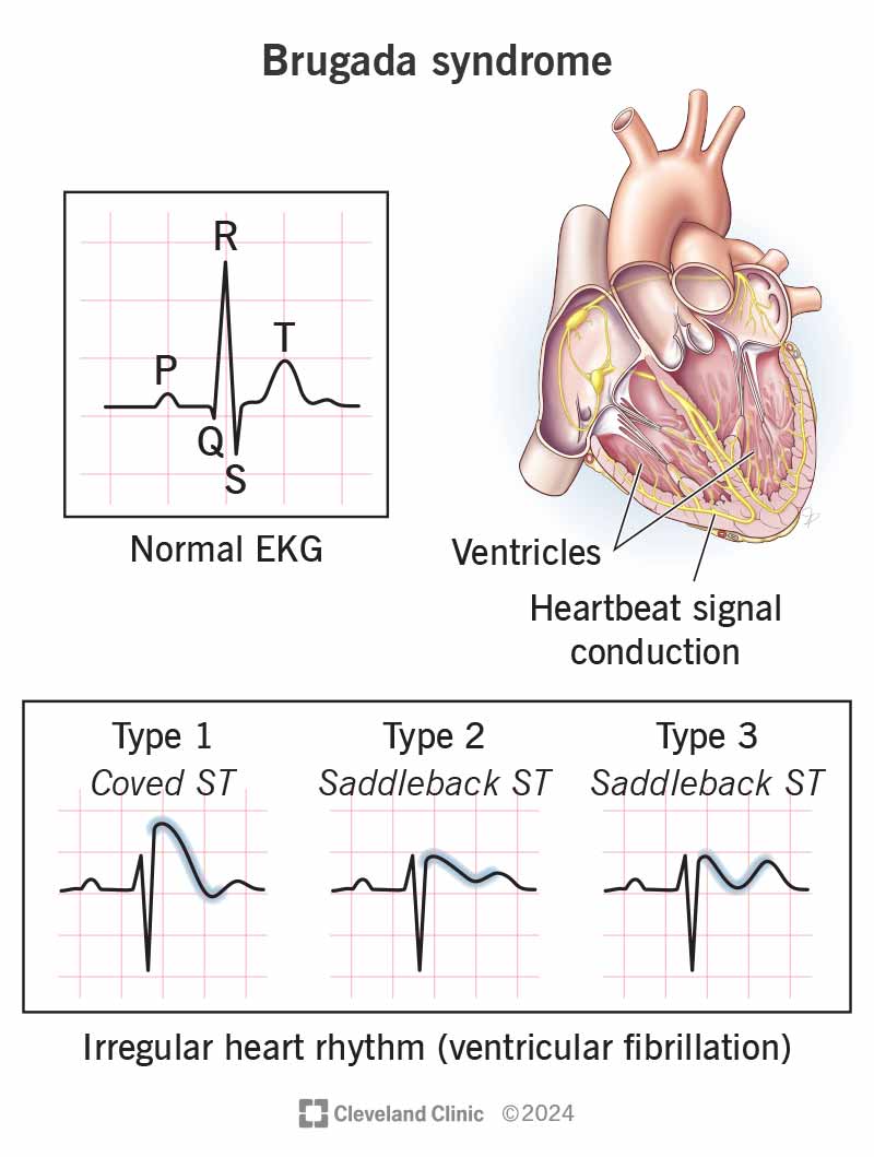
Brugada syndrome

Brugada syndrome

Genetic disorderWikipedia

Search complete. 18 mentions across 8 episodes found for "Brugada syndrome".

Oct 27, 2026

Kamoa TamarisaGUEST
23:47
But the important thing to remember is natrolol is highly secreted in the breast milk.
Kamoa TamarisaGUEST
23:53
And moving on to Brugada syndrome.
Kamoa TamarisaGUEST
23:54
Brugada syndrome, the recommendations were based upon 2023 document.
Kamoa TamarisaGUEST
24:00
And what's key in Brugada is telemetry is important.
Kamoa TamarisaGUEST
24:04
Placing the defibrillator pads, especially if the patient is having any runs of salvos of VT.

Unknown podcast

ECG Pitfalls — Mistakes in Interpretation and How to Record an ECG in the OPD

Oct 11 · 5 Mentions

A M ThirugnanamGUEST
9:31
When there's ST depression in V1 to V3 or a strong suspicion of a circumflex occlusion with a non-diagnostic ECG, record the posterior leads V7, V8, and V9.
A M ThirugnanamGUEST
9:47
When Brugada syndrome is suspected, recording V1 and V2 one or two intercostal spaces higher, in the second or third space, increases sensitivity.
A M ThirugnanamGUEST
10:00
And when you need to see the atrial activity clearly, for example, to find hidden P waves in a broad complex tachycardia, the Lewis lead, with the electrodes placed to look at the atria, can help.
A M ThirugnanamGUEST
10:15
Each of these takes only a minute.
A M ThirugnanamGUEST
11:49
The commonest is placing V1 and V2 too high, in the second or third intercostal space.
A M ThirugnanamGUEST
11:57
That produces an RSR' pattern that looks like incomplete right bundle branch block: P waves that become negative or biphasic in V1 and V2 and sometimes T wave inversion.
A M ThirugnanamGUEST
12:12
It can also mimic a Brugada pattern or a septal infarction.
A M ThirugnanamGUEST
12:17
A clue that the leads are too high is a negative P wave in V2, because from above, the lead is looking down at the atria.

Unknown podcast

Benign and Malignant Arrhythmias: Diagnosis, Treatment and Management

Oct 11 · 3 Mentions

A M ThirugnanamGUEST
28:48
And screening of first-degree relatives is essential.
Alexander WoodHOST
28:53
What about Brugada syndrome? It's often described as particularly relevant in Asia.
A M ThirugnanamGUEST
29:01
Yes.
A M ThirugnanamGUEST
29:02
Brugada syndrome is more prevalent in South East Asia and is an important cause of sudden death in young men, typically during sleep.
A M ThirugnanamGUEST
29:12
It is linked in some families to SCN5A mutations.
A M ThirugnanamGUEST
29:16
The diagnostic pattern is the type 1 Bru-gade ECG.
A M ThirugnanamGUEST
29:32
The leads can be placed one or two intercostal spaces higher to unmask it.
A M ThirugnanamGUEST
29:38
The pattern may be intermittent and can be provoked by fever or by a sodium channel blocker challenge, such as a gimilinoflicanide performed in a monitored setting.

Unknown podcast

The Missed Diagnosis: Why We Miss Important Diagnoses and How to Fix the Blunder

Oct 11 · 3 Mentions

A M ThirugnanamGUEST
12:58
Wellens' syndrome, deep or biphasic T waves in V2 and V3 in a pain-free patient signals a critical proximal LAD stenosis.
A M ThirugnanamGUEST
13:10
Other important missed findings include a raised serum potassium, Brugada pattern, Wolff-Parkinson-White syndrome, a long QT interval, and hypertrophic cardiomyopathy with deep T wave inversion.
A M ThirugnanamGUEST
13:26
And a single normal electrocardiogram never excludes an acute coronary syndrome.
A M ThirugnanamGUEST
13:33
Repeat it.
A M ThirugnanamGUEST
13:45
Most episodes are indeed reflex or vasovagal, but syncope can be the only warning before sudden cardiac death.
A M ThirugnanamGUEST
13:54
Red flags include syncope during exertion or while lying down, syncope preceded by palpitations or with no warning, syncope with chest pain or breathlessness, a family history of sudden death below 50, known structural heart disease, and abnormal electrocardiogram.
A M ThirugnanamGUEST
14:17
Causes we mustn't miss include severe aortic stenosis, hypertrophic cardiomyopathy, complete heart block or high grade atrioventricular block, ventricular tachycardia, long QT syndrome, Brugada syndrome, pulmonary embolism, aortic dissection, and in the elderly, internal bleeding.
A M ThirugnanamGUEST
14:42
So every patient with syncope deserves a careful history, examination, lying and standing blood pressure, and an electrocardiogram, and when red flags are present, an echocardiogram and rhythm monitoring.

Unknown podcast

Cardiac Arrhythmias — Benign and Malignant, How to Diagnose, Acute Treatment and Chronic Care

Oct 11 · 2 Mentions

A M ThirugnanamGUEST
7:45
High grade AV block, Mobitz type 2 and complete heart block, which can cause syncope and asystole.
A M ThirugnanamGUEST
7:53
And the arrhythmias of inherited conditions: long QT syndrome, Brugada syndrome, catecholaminergic polymorphic ventricular tachycardia, arrhythmogenic cardiomyopathy, and hypertrophic cardiomyopathy, which may present for the first time as sudden death in a young person.
SamanthaHOST
8:17
Let's turn to diagnosis.
SamanthaHOST
8:20
Where does it start?

25 MINS LATER

A M ThirugnanamGUEST
32:59
Correct electrolytes promptly during illness.
A M ThirugnanamGUEST
33:03
High-risk patients may need a defibrillator or left cardiac sympathetic denervation.
A M ThirugnanamGUEST
33:10
In Brugada syndrome, the key lifestyle measures are prompt treatment of fever with paracetamol because fever can trigger arrhythmias and avoidance of certain drugs.
A M ThirugnanamGUEST
33:23
The Brugada Drugs website lists them.

Unknown podcast

The Secrets Never Told During Internship: Expert Diagnosis in Cardiology

Oct 11 · 1 Mention

A M ThirugnanamGUEST
20:17
Look for De Winter T waves and Wellens pattern in the anterior leads, which indicate a proximal LAD problem.
A M ThirugnanamGUEST
20:27
In a patient with syncope, look for a short PR interval and a delta wave, a long QT interval, a Brugada pattern in V1 and V2, bifascicular block, or an epsilon wave.
A M ThirugnanamGUEST
20:42
In a breathless patient, look for sinus tachycardia with a right bundle branch block, right axis deviation, and T wave inversion in V1 to V4 together with the inferior leads, which suggests right ventricular strain from pulmonary embolism.
A M ThirugnanamGUEST
21:05
In an elderly patient with heart failure, low voltage with thick walls on echo suggests amyloidosis.

Unknown podcast

Facing Cardiac Emergencies — Ensuring Safety, Quality of Care and Saving Patients' Lives

Oct 11 · 1 Mention

A M ThirugnanamGUEST
22:25
Most syncope is benign and reflex mediated, but some is the first sign of a lethal arrhythmia.
A M ThirugnanamGUEST
22:33
The red flags are syncope during exertion or while lying down, syncope without warning, palpitations just before the event, a family history of sudden death, non-structural heart disease, and an abnormal ECG, for example, Mobitz type 2 or complete heart block, a long or short QT, a Brugada pattern, pre-excitation, or signs of cardiomyopathy.
A M ThirugnanamGUEST
23:02
Patients with these features should be monitored and investigated before discharge, not sent home with reassurance.
SamanthaHOST
23:11
Bleeding is a frequent complication of cardiac treatment.
Patil ArmenianHOST
9:46
But there are aberrancies which are kind of slowdowns, um, in conduction due to a lot of different things.
Patil ArmenianHOST
9:52
And so that's, for example, your Brugada syndromes, AV dissociations, fusion beats, all these other things that honestly are interesting academically, but in real life they don't really matter, um, whether in the, you're in the pre-hospital or hospital setting, frankly, to manage a really sick, unstable person.
Saajan BhaktaHOST
10:12
Right.
Saajan BhaktaHOST
10:13
So just as Patil was saying, not every wide complex tachycardia is V-tach.

We value your privacy

We use cookies to understand how you use our platform and to improve your experience. Click “Accept All” to consent, or “Decline non-essential” to opt out of non-essential cookies. Read our Privacy Policy.